Pages

Showing posts with label glia. Show all posts

Tuesday, June 18, 2013

Can Human Brain Tissue Make Mice Smarter? Emory Neuroethics Journal Club Review

What makes humans smart?  This was the primary question posed in the final Journal Club of the Spring 2013 semester.  Led by Riley Zeller-Townson, the club discussed Han et al. (2013), a paper that discusses the enhancement of learning in mice after grafting human glial progenitor cells into their brains. Riley began by explaining the paper and the work leading up to it. Most of the roles of glial cells involve supporting and protecting neurons, such as synaptic plasticity, myelination, and maintaining the blood-brain barrier (Barres, 2003). This study focuses on one subtype of glia, called astrocytes, cells that provide nutrients to neurons (Tsacopoulos et al, 1996).


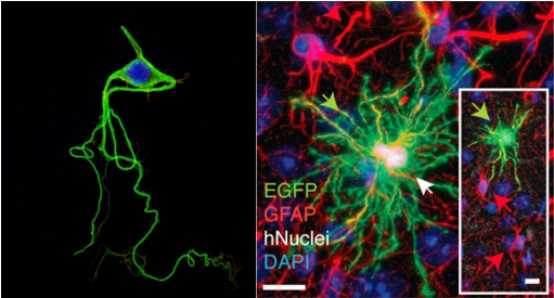
Neurons (shown on left) possess both axons and dendrites and are shaped differently than glial cells (Source).  The glial cell shown on the right is an astrocyte, which is more “star” shaped due to its many branched processes (Han et al 2013).

While people generally think of neurons as being the important type of brain cells, research is beginning to show that the merits of glial cells were previously underestimated. Interestingly, post-mortem analysis of Albert Einstein’s brain showed that he had more glia than the average person (Diamond et al, 1985).  Along the same vein, previous studies have shown that primate glia are larger, more complex, and faster than those of mice (Colombo, 1996; Oberheim et al., 2009). Therefore, is it possible that glial cells are the root of intelligence?

Han et al. tested this hypothesis by grafting human astroglia progenitors into neonatal immune-deficient mice.  Because the mice retained their own astroglia as well, the mice were chimeric, or having both mouse and human glia.  The human glial cells were fluorescently labeled before implantation so that they could be identified after each mouse was sacrificed, which occurred anywhere from 0.5-20 months of age.  The successfully grafted glial cells were found to have distributed across the forebrain, including the hippocampus and cortex.  Additionally, human glia were found in the amygdala, thalamus, and neostriatum in mice ages 12-20 months.


Next, the authors did a battery of tests that revealed that the cells were not only present, but also functional.  The authors found that not only were the human astrocyte progenitor cells present in the mouse brain, but they actually differentiated into humanoid protoplasmic astrocytes that formed synapses with mouse astrocytes.  In the chimeric mouse brain, the human astrocytes produced Ca2+ signals that were three times faster than Ca2+ signals produced by mouse astrocytes (this had previously been demonstrated in human tissue, but held true for chimeric mouse tissue). Next, the authors compared hippocampal dentate synaptic activity in chimeric mice to unengrafted and allografted mice and found that tissues with engrafted human glia showed a greater level of excitatory synaptic transmission. This finding goes hand in hand with the next subsequent finding that Long Term Potentiation (LTP), an experimental measure correlated with memory formation, increased in chimeric mice. The authors speculate that this increase was due to insertion of GluR1, a specific type of excitatory receptor often associated with memory and learning, into mice neurons by human glia, which lowered the threshold for LTP induction.


Because LTP was increased in chimeric mice, one would assume that these mice, not just their cells, would also showed quicker learning behaviors than the control mice. As expected, mice engrafted with human glia performed better in all four behavioral tasks compared to unengrafted and allografted mice, showing that the improvement was due to the human glia, not the act of engrafting cells into the mice.


Overall, the results of the paper suggest that astrocytes play a role in learning, and that more complex astrocytes might increase learning by lowering the threshold at which LTP occurs. Furthermore, the paper implies that human astrocytes, and possibly other subtypes of untested human glial cells, play a role in the high level of cognition observed in humans (and possibly in mice).


The Rats of
NIMH (Source)


After introducing the paper and its findings, the subject turned to the ethical considerations associated with the study.  Riley introduced another paper (Greely et al 2007) as a response to Han et al.  In his paper, Greely discusses the possible ethical issues that would arise by performing this type of study.  Greely lists several possible ethical issues, including the risk of conferring humanity upon the mice, the potential for pain and suffering, public reaction, and respect for human tissues.


In response to Greely’s first concern, journal club attendee and bioethics professor Dr. Jonathan Crane pointed out that “conferring humanity” upon the mice was the wrong term, as “humanity” simply means the traits that make us human.   Dr. Crane pointed out that humans do not really have any unique capabilities that other animals do not have, we just have them in a different degree.  He points out the Greely is most likely concerned about the risk of conferring “personhood,” or making the mouse a autonomous individual to the same extent that an adult human is.


Of course, if personhood were conferred upon a species that we cannot communicate with, would we even be able to recognize it?  It seems a big issue with this type of study is not only the possibly innately unethical problem of creating an unnatural sentient being, but also the issue of creating a (more?) autonomous being, not realizing it as such, and continuing to treat it like a typical lab animal.


Greely’s second concern, the potential for pain and suffering in the chimeric mouse, was also addressed.  While Greely’s paper noted the possibility of increased pain in the mouse due to having human neurons, the group discussed the mouse’s possibly increased emotional pain as well.  It was addressed that a mouse with human “intelligence” might also gain a human-like propensity for depression or realizing the futility of life as an experimental animal.   The authors seem to have guarded against this in the Han et al. study by noting that the chimeric mice were just as social as normal mice.  However, if these types of studies continue and mice intelligence is further increased, depression and suffering in the chimeric mouse will be a concern. The experimenters compared the reaction times and pain thresholds of chimeric mice to normal mice and found them to be the same.  These findings, coupled with the observation that the chimeric mice are just as social as normal mice are checks the experimenters used to show that the mice were not suffering mentally or physically.  These checks should also be present in future studies with chimeric species.  However, the downside is that these tests cannot gauge suffering until it has already happened.


Greely’s concern about public reactions was also discussed.  When discussing public reactions, it was addressed that different groups would be opposed to this study for different reasons.  Some groups would be opposed because of the belief that implanting human brain cells into a disvalues human intelligence.   Emory medical ethicist Dr. John Banja noted that some would question whether or not it is ethical to create hybrid species in the first place.  Dr. Crane pointed out that the study is also an affront to the mouse, as it is attempting to improve an animal that is perfect to begin with.


Riley’s main concern was that progression of this experiment will continue to create new species that we know nothing about.  Without knowing which mouse traits and which human traits a hybrid will have, it is impossible to care for it in the correct way from the beginning.  Riley pointed out that sometimes it takes thousands of years to create a cultural consensus on what is ethical regarding a certain issue, so if a new species pops up overnight society may not treat the animals respectfully.  Furthermore, these animals could completely change biological science: furthering this study could lead to a greater understanding of the mechanisms for learning, memory, and eventually even consciousness in humans.  An alternative to this however would be is a unique and species is created that is later discovered to have human-like consciousness and increased suffering.  The scientific community would stall as it would have to deal with public dissent amidst trying to find a more ethical method of testing the same hypotheses.


While future advancements along this line of work could have the potential to help us understand exactly what intelligence is, how it works, and why some people have more of it than others, these types of experiments could also have the ability to create a brand new species that society is not ready to care for or treat with respect.  Furthermore, it may not be possible to determine when the ethical boundary has been overstepped until it has already happened. Therefore, in continuations of this study, it is important to use chimeric animals with discretion and continue testing for possible suffering.


If you missed out on this journal club meeting you can watch a video of it and previous meetings here.


References


Alexander V. Gourine, V. K. (2010). Astrocytes Control Breathing Through pH-Dependent Release of ATP. Science, 571-575.


Barres, B. A. (2003). What is a Glial Cell? Glia, 4-5.


Colombo, J. (1996). Interlaminar astroglial processes in the cerebral cortex of adult monkeys but not of adult rats. Cells Tissues Organ, 57-62.


Henry T. Greely, M. K. (2007). Thinking About the Human Neuron Mouse. The American Journal of Bioethics, 27-40.


Magistretti, M. T. (1996). Metabolic Coupling between Glia and Neurons . Journal of Neuroscience, 877-885.


Marian C. Diamond, A. B. (1985). On the brain of a scientist: Albert Einstein. Experimental Neurology, 198–204.


Oberheim, N. W. (2006). Astrocytic complexity distinguishes the human brain. Trend in Neuroscience, 547-553.


Xiaoning Han, M. C. (2013). Forebrain Engraftment by Human Glial Progenitor Cells Enhances Synaptic Plasticity and Learning in Adult Mice. Cell, 342-353.


Want to cite this post?


Young, E. (2013). Can Human Brain Tissue Make Mice Smarter? Emory Neuroethics Journal Club Review. The Neuroethics Blog. Retrieved on

, from http://www.theneuroethicsblog.com/2013/06/can-human-brain-tissue-make-mice.html.

Friday, May 24, 2013

Now Available! Neuroethics Journal Club Video Archives on YouTube


The Neuroethics Journal Club videos are now available on YouTube. Watch each discussion to learn about a variety of neuroethics issues, from treatments for pedophilia to neural plasticity in mice. For each video, one presenter introduced the journal topic and opened discussion to the audience. 


Neuroethics Journal Club: The Sexed Brain


The Sexed Brain: Between Science and Ideology


Catherine Vidal, Neuroethics, 2012 


Abstract: Despite tremendous advances in neuroscience, the topic “brain, sex and gender” remains a matter of misleading interpretations, that go well beyond the bounds of science. In the 19th century, the difference in brain sizes was a major argument to explain the hierarchy between men and women, and was supposed to reflect innate differences in mental capacity. Nowadays, our understanding of the human brain has progressed dramatically with the demonstration of cerebral plasticity. The new brain imaging techniques have revealed the role of the environment in continually re-shaping our brain all along our lifetimes as it goes through new experiences and acquires new knowledge. However, the idea that biology is a major determining factor for cognition and behavioral gender differentiation, is still very much alive. The media are far from being the only guilty party. Some scientific circles actively promote the idea of an innate origin of a gender difference in mental capacities. Experimental data from brain imaging, cognitive tests or genetics are often distorted to serve deterministic ideas. Such abuse of “scientific discourses” have to be counteracted by effective communication of clear and unbiased information to the citizens. This paper presents a critical analysis of selected examples which emphasize sex differences in three fields e.g. skills in language and mathematics, testosterone and financial risk-taking behavior, moral cognition. To shed light on the data and the methods used in some papers, we can now—with today’s knowledge on cerebral plasticity—challenge even more strongly, many false interpretations. Our goal here is double: we want to provide evidence against archaic beliefs about the biological determinism of sex differences but also promote a positive image of scientific research.


Neuroethics Journal Club: Pedophilia


Real-time functional magnetic imaging—brain–computer interface and virtual reality: Promising tools for the treatment of pedophilia 


Renaud et al, 2011


Abstract: This chapter proposes a prospective view on using a real-time functional magnetic imaging (rt-fMRI) brain-computer interface (BCI) application as a new treatment for pedophilia. Neurofeedback mediated by interactive virtual stimuli is presented as the key process in this new BCI application. Results on the diagnostic discriminant power of virtual characters depicting sexual stimuli relevant to pedophilia are given. Finally, practical and ethical implications are briefly addressed.


Neuroethics Journal Club: Social Pain 


The pain of social disconnection: examining the shared neural underpinnings of physical and social pain 


Eisenberger, 2011 


Nature Reviews Neuroscience


Abstract: Experiences of social rejection, exclusion or loss are generally considered to be some of the most 'painful' experiences that we endure. Indeed, many of us go to great lengths to avoid situations that may engender these experiences (such as public speaking). Why is it that these negative social experiences have such a profound effect on our emotional well-being? Emerging evidence suggests that experiences of social pain--the painful feelings associated with social disconnection--rely on some of the same neurobiological substrates that underlie experiences of physical pain. Understanding the ways in which physical and social pain overlap may provide new insights into the surprising relationship between these two types of experiences.


Neuroethics Journal Club: Plasticity and Learning 


Forebrain Engraftment by Human Glial Progenitor Cells Enhances Synaptic Plasticity and Learning in Adult Mice 


Han et al, 2013 Cell Stem Cell


Abstract: Human astrocytes are larger and more complex than those of infraprimate mammals, suggesting that their role in neural processing has expanded with evolution. To assess the cell-autonomous and species-selective properties of human glia, we engrafted human glial progenitor cells (GPCs) into neonatal immunodeficient mice. Upon maturation, the recipient brains exhibited large numbers and high proportions of both human glial progenitors and astrocytes. The engrafted human glia were gap-junction-coupled to host astroglia, yet retained the size and pleomorphism of hominid astroglia, and propagated Ca2+ signals 3-fold faster than their hosts. Long-term potentiation (LTP) was sharply enhanced in the human glial chimeric mice, as was their learning, as assessed by Barnes maze navigation, object-location memory, and both contextual and tone fear conditioning. Mice allografted with murine GPCs showed no enhancement of either LTP or learning. These findings indicate that human glia differentially enhance both activity-dependent plasticity and learning in mice.


Stay tuned for more Neuroethics Journal Club videos next year. Also, to get daily updates about emerging ideas in neuroethics, follow Emory Neuroethics on Facebook here and Twitter here.


Thursday, August 2, 2012

A Case Study in Misunderstanding

The media gets a bad rap for oversimplifying, misrepresenting, and even sensationalizing stories. Stories about science are no exception. But it is also no secret that the scientists themselves are under all sorts of pressures to oversimplify, misrepresent, and even sensationalize the science.


But sometimes even good science and good reporting can lead to inaccuracies in “translation” that may look like (intentional) oversimplification, misrepresentation, or even sensationalization, when in fact these inaccuracies are due to honest, understandable, and avoidable misunderstandings.


One of the sources of this honest misunderstanding comes from cognitive neuroscience’s usage of everyday mental terms. Often the scientists’ usage of mental terms differ in important respects from the everyday understanding of these mental terms, and the particular idiosyncratic (from the everyday-use point of view) ways in which the scientists are employing the terms aren’t always made explicit.[1]


For an example of how the misunderstanding in use of mental terms can lead to what may look like sensationalized reporting, see the numerous media reports [2][3][4][5]on Han and colleagues [6] finding that acute cannabinoids impair working memory in mice through glial cell receptors.


Glowing lab mice. True story. But different experiment.


According to the media reports, the scientists uncovered a surprising cellular mechanism—glia, not neurons!—for working memory deficits associated with marijuana use. So far, so good: This is what the scientists claimed they were testing in their study and is also what they claimed to have found in their study.


But confusion quickly arises.


Not too far into a Scientific American article by Ruth Williams[2], the author notes, "This is one of the first studies to suggest that glia play a key role in conscious thought." And a Big Think media report [3] is even titled "Marijuana Opens New Field in Consciousness Research."


Whoa! Mice have now become a model to study conscious thought! Amazing! How in the world do the media reporters go from "glia plays a role in marijuana-related memory deficits in mice" to "consciousness research"?


Most people's first reaction is probably: This must be a case of media sensationalization. But I must admit my first reaction was: This must be a case of scientist sensationalization (and the media is merely passing the message along).


But the more likely explanation is probably a lot less sinister; it is probably a case of honest, understandable, and avoidable misunderstandings.


When the reporter hears the term ‘working memory’, she thinks about working memory in the same way most reasonable and educated people would. For example, in the Scientific American article, Ruth Williams [2] defined working memory as “the short-term memory we use to hold on to and process thoughts.” And this idea of “holding on to and processing thoughts” in working memory is usually thought of as a conscious, explicit, and often effortful process. (When I think about working memory, I naturally think of working a complex math problem in my head or the effortful rehearsing of a phone number.)


This line of thinking could quickly lead one to (reasonably) believe that the researchers have uncovered a cellular mechanism associated with effortful conscious thought. And, of course, as noted above, the reporters do end up making such claims.


Unfortunately, Han and colleagues aren’t talking about this sort of conscious, explicit, and effortful working memory processes. Rather, they are talking about spatial working memory, and, in particular, spatial working memory processes that are not necessarily explicit, conscious, or effortful. Even more unfortunately, these types of working memory processes may have cellular mechanisms that are dissociable from the more commonly-associated-with-working-memory conscious, explicit, and effortful processes.


And even though Han and colleagues are clear in their article that they are talking about spatial working memory, they are not clear that they are talking about processes that are not necessarily explicit, conscious, or effortful. Additionally, Han and colleagues are not particularly clear in communicating the idea that the findings associated with deficits in non-explicit, non-conscious, non-effortful spatial working memory processes may have little to nothing to do with the sorts of working memory processes that would typically come to mind when a reasonable and educated person was to think of working memory.


Han and colleagues are assuming Badley's Working Memory Model. Consciousness not assumed. (Especially for the visuo-spatial scratchpad and the phonological loop.)


Thus, the “consciousness” claims made by the media are probably not the result of media sensationalism. Rather, these claims are more likely the result of honest, understandable, and avoidable misunderstandings.


And these misunderstanding may actually matter.


For example, Han and colleague motivate their study by talking about the potential therapeutic benefits of marijuana. In the opening paragraph of the journal article, they note that “the potential therapeutic use of marijuana is limited by important side-effects associated with its use,” especially “the impairments of working memory in humans.” One of the implications being that if we could understand the cellular mechanisms of marijuana-induced working-memory impairments, maybe we could make a drug that could give the therapeutic benefits of marijuana without causing impairments in working memory. [7]


And it seems that Han and colleagues findings suggest just this potential. As noted by Williams [2], “Unlike THC’s effect on memory, its pain-relieving property appears to work through neurons. In theory, therefore, it might be possible to design THC-type drugs that target neurons—but not glia—and offer pain relief without the forgetfulness.”


This sounds like an exciting potential, but the potential is really only exciting to the extent that one misreads the implications of Han and colleagues findings. Han and colleagues findings shed light on a very particular working-memory process, and the findings related to this particular working-memory process may not have much to do with the sort of marijuana-induced memory deficits that we care the most about.


Want to cite this post?


Shepard, J. (2012). A Case Study in Misunderstanding. The Neuroethics Blog. Retrieved on
, from http://www.theneuroethicsblog.com/2012/08/a-case-study-in-misunderstanding.html


[1] Figdor, C. (forthcoming) What is the “cognitive” in cognitive neuroscience?” Neuroethics.

[2] http://www.scientificamerican.com/article.cfm?id=marijuana-reveals-memory-mechanism

[3] http://bigthink.com/ideafeed/marijuana-opens-new-field-of-consciousness-research

[4] http://www.rawstory.com/rs/2012/07/13/stoned-mice-lead-to-breakthrough-in-understanding-memory/

[5] http://www.tokeofthetown.com/2012/07/how_marijuana_affects_memory_its_not_the_neurons.php

[6] Han, J., Kesner, P., Matna-Laurent, M., Duan, T. Xu, L., Georges, F., Koehl, M., Abrous, D., Mendixabal-Zubiaga, J., Grandes, P., Liu, Q., Bai, G., Wang, W., Xiong, L., Ren, W., Marsicano, G., & Zhang, X. (2012) Acute cannabinouds impair working memory through astroglial CB1 receptor modulation of hippocampal LTD. Cell, 5, 1039-1050.

[7] In my opinion, Han and colleagues actually welcome the misunderstanding by framing their study in this way. After all, when we think about marijuana-induced forgetfulness (or even more to the point, the most salient problems associated with marijuana-induced forgetfulness), it does not seem that we are primarily concerned with the particular phenomenon that is actually under study in Han and colleagues study.