Pages

Showing posts with label mice. Show all posts

Tuesday, November 17, 2015

Do you have a mouse brain? The ethical imperative to use non-human primates in neuroscience research

by Carlie Hoffman


Much of today’s neuroscience research investigating human brain diseases and disorders utilizes animal models. Animals ranging from flies, rodents, and non-human primates are routinely used to model various disorders, with mice being most commonly utilized. Scientists employ these animal models to approximate human conditions and disorders in an accessible manner, with the ultimate purpose of applying the findings derived in the animal back into the human brain.


Rhesus macaques, a species of NHP often used in research.


The use of animals in research has been the source of much debate, with people either supporting or objecting their use, and objections arising from animal rights activists, proponents of critical neuroscience such as Nikolas Rose and Joelle Abi-Rached, and others. A main focus of this debate has also been the use of non-human primates (NHP) in research. The cognitive functions and behaviors of NHPs are more closely related to those seen in humans than are rodent cognitions and behaviors, thus causing primates to be held as the closest approximation of human brain functioning in both normal and disease states. Though some say NHP research is essential, others call for scaling down or even completely eliminating it. Strides have already been made towards the reduction and removal of NHPs from experimental research, as displayed by the substantial justification required to perform experiments utilizing them, the increasing efforts going towards developing alternative non-animal models (including the Human Brain Project’s goal to create a computer model of the human brain), and the recent reduction of the use of chimpanzees in research [2, 6].  A case was even brought to the New York Supreme Court earlier this year to grant personhood status to two research chimpanzees.


However, if NHPs are completely removed from human brain disease research, this leaves rodents as the primary non-human animal model for the human condition. This raises an important question: are we (both the general public and scientists) okay with accepting a detailed understanding of the mouse brain as being equivalent to a detailed understanding of the human brain? Dr. Yoland Smith, a world-renowned Emory researcher in neurodegenerative disease, says no— work with both NHPs and rodent models must continue to complement and inform each other.


Smith came to Emory University in 1996 and currently works at the Emory-affiliated Yerkes National Primate Research Center. Smith’s work is 90% rhesus macaque-based and 10% rodent-based, with the aim of most of his rodent work being to fine-tune new approaches and methods to apply to his rhesus macaques. While many animal rights advocates feel it is an ethical imperative to eliminate NHP research, Dr. Smith believes there are currently more pressing obstacles to the continued use of NHPs to study neuroscience, namely the increasing regulatory constraints on NHP research and the challenges in translating rapid technology development from mice to NHPs. 


Mice are the animals most commonly used in research.


For instance, the number of rodents (almost exclusively mice) utilized in biomedical research has been increasing at a dramatic pace compared with that of primates, the latter accounting for only about 0.1- 0.3% of all animals currently used in biomedical research in the United States and Europe. This is in part due to the ease of acquiring and maintaining rodent colonies as compared to NHP colonies: the cost of maintaining a mouse at Emory ranges from $0.83- $4.13 per day, while the cost of maintaining a NHP is on the range of $80-$110 per day [3]. While ultimately rodent research is not cheap (most researchers maintain rodent colonies containing anywhere from tens to hundreds of mice, causing total per diem costs to rise rapidly), the relatively lower cost of maintenance and ease of access affiliated with rodent use have allowed for extensive experimental optimization and exploratory testing to be performed in rodents, but not in NHPs. As a result, there has been advancement and development of rodent-based technologies over NHP-based technologies, and techniques such as in vitro electrophysiology, optogenetics, and transgenic strains have been developed for use in mice. Because such techniques do not cross easily into NHP research, they are not currently available for use in the NHP brain. Consequently, new methods are continually being applied and developed almost exclusively for use in the mouse, while the needed time and money to adapt these assays to NHPs are not being spent. Scientists are thus able to dig deeper and answer more sophisticated questions about the mouse brain, while NHP research is being left in the dust. NHP research may ultimately come to an end not just because of the ethical arguments against it, but because rodent research is leaps and bounds ahead of it.


This trajectory also raises questions about what role we think NHP research should continue to play in the field of neuroscience: Can NHP research ever match the pace of rodent research? Should we use NHPs at all? Can all of our questions about the human brain and human health be answered using rodent models?


Smith’s answer to this final question is no. Although Smith feels rodent research produces highly valuable information, contributes to advances in neuroscience, and must continue to grow at a fast pace, he posits that it would be naïve to believe that gaining knowledge about the mouse brain is sufficient to achieve our ultimate goal of “getting to the human.” While it would be easy to say that the human brain is equivalent to the mouse brain, he states this is simply not true—particularly when examining complex brain functions and diseases that involve high-order cortical processing. For instance, rodents are often used to model many complex neuropsychiatric diseases affecting the prefrontal cortex and influenced by social and cultural components; however, there are striking differences in the size and complexity of the prefrontal cortex and other associative cortices in primates versus rodents, and animal models are unable to replicate the integral role culture plays in psychiatric disease pathology (a topic also described in a previous blog post) [1, 4]. The overly simplistic organization of the rodent cerebral cortex compared with that of humans, Dr. Smith believes, makes NHP research absolutely essential. If we hope to make significant progress toward the development of new therapeutic strategies for complex cognitive and psychiatric disorders, then there is an urgent need for the ongoing development of NHP models of such prevalent diseases of the human brain.


Comparative images of human, mouse, and rhesus brains.


Smith also believes there is a critical knowledge gap between the rodent brain and the human brain that cannot be addressed without a deeper understanding of both the healthy and diseased NHP brain. NHP research has already partially helped to fill this gap [5], as evidenced by the breakthroughs made in understanding disease pathophysiology and the development of surgical therapies for Parkinson’s disease through the use of NHP models, the focus of Smith and his colleagues’ work at Emory. However, Smith notes that these advances would not have been possible without a strong foundation of rodent research. This interplay between primate and rodent work serves as a model for how Smith hopes the field of neuroscience will progress in the future, with both rodent research and primate research growing together in parallel and feeding into one another, ultimately helping researchers to “get to the human.”


If significant strategies are not put in place by funding agencies to maintain continued support for NHP research, this parallel growth will not be possible. Smith urges that we maintain NHP research by continuing to develop technologies for use in NHPs and by generating discussions about the use and drawbacks of NHPs in neuroscience research. Smith feels that strides toward maintaining NHP research should be spearheaded by larger funding institutions, such as the National Institutes of Health (NIH). He proposes there should be a call for grant applications specifically involving NHP research as well as the formation of an NIH-based committee to discuss how we can perform NHP research in a successful way; to assess whether we, as a scientific community, are always moving toward the human; and to examine the ethical and practical limitations of using NHPs in research. Smith also encourages scientists to be proactive and unafraid to engage the community in discussions of the ethical dilemmas surrounding primate research—we cannot ignore these issues and instead must be the mediators of such ethical discussions.


Dr. Smith believes NHP research is essential to advancing the understanding of the human brain and improving human health. As such, Smith does not think that this research will ever fully disappear. While young researchers might find the current funding climate and research constraints daunting, he claims it is the responsibility of current primate researchers to continue to train new scientists; if we do not train new NHP scientists, we will just be contributing to the loss. Therefore, while NHP research is receiving pressure both from society and from scientists, the use of primates in neuroscience research must continue. After all, as Smith stated, “I do not have a mouse brain.”


Works Cited


1. Ding, SL (2013) Comparative anatomy of the prosubiculum, subiculum, presubiculum, postsubiculum, and parasubiculum in human, monkey, and rodent. J Comp Neurol 521: 4145-4162. doi: 10.1002/cne.23416


2. Doke, SK, & Dhawale, SC (2015) Alternatives to animal testing: A review. Saudi Pharmaceutical Journal 23: 223-229. doi: http://dx.doi.org/10.1016/j.jsps.2013.11.002


3. International Animal Research Regulations: Impact on Neuroscience Research: Workshop Summary. (2012). Washington DC: National Academy of Sciences.


4. Nestler, EJ, & Hyman, SE (2010) Animal models of neuropsychiatric disorders. Nat Neurosci 13: 1161-1169. doi: 10.1038/nn.2647


5. Phillips, KA, Bales, KL, Capitanio, JP, Conley, A, Czoty, PW, t Hart, BA, Hopkins, WD, Hu, SL, Miller, LA, Nader, MA, Nathanielsz, PW, Rogers, J, Shively, CA, & Voytko, ML (2014) Why primate models matter. Am J Primatol 76: 801-827. doi: 10.1002/ajp.22281


6. Tardif, SD, Coleman, K, Hobbs, TR, & Lutz, C (2013) IACUC review of nonhuman primate research. ILAR J 54: 234-245. doi: 10.1093/ilar/ilt040


Want to cite this post?


Hoffman, C. (2015). Do you have a mouse brain? The ethical imperative to use non-human primates in neuroscience research. The Neuroethics Blog. Retrieved on , from http://www.theneuroethicsblog.com/2015/11/do-you-have-mouse-brain-ethical.html

Tuesday, June 18, 2013

Can Human Brain Tissue Make Mice Smarter? Emory Neuroethics Journal Club Review

What makes humans smart?  This was the primary question posed in the final Journal Club of the Spring 2013 semester.  Led by Riley Zeller-Townson, the club discussed Han et al. (2013), a paper that discusses the enhancement of learning in mice after grafting human glial progenitor cells into their brains. Riley began by explaining the paper and the work leading up to it. Most of the roles of glial cells involve supporting and protecting neurons, such as synaptic plasticity, myelination, and maintaining the blood-brain barrier (Barres, 2003). This study focuses on one subtype of glia, called astrocytes, cells that provide nutrients to neurons (Tsacopoulos et al, 1996).


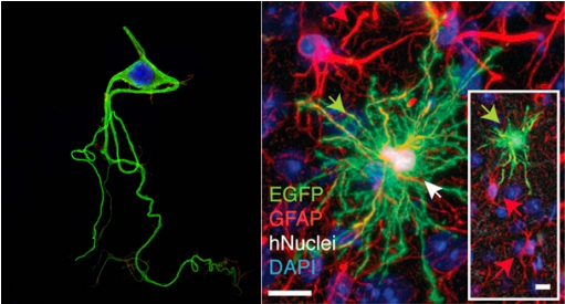
Neurons (shown on left) possess both axons and dendrites and are shaped differently than glial cells (Source).  The glial cell shown on the right is an astrocyte, which is more “star” shaped due to its many branched processes (Han et al 2013).

While people generally think of neurons as being the important type of brain cells, research is beginning to show that the merits of glial cells were previously underestimated. Interestingly, post-mortem analysis of Albert Einstein’s brain showed that he had more glia than the average person (Diamond et al, 1985).  Along the same vein, previous studies have shown that primate glia are larger, more complex, and faster than those of mice (Colombo, 1996; Oberheim et al., 2009). Therefore, is it possible that glial cells are the root of intelligence?

Han et al. tested this hypothesis by grafting human astroglia progenitors into neonatal immune-deficient mice.  Because the mice retained their own astroglia as well, the mice were chimeric, or having both mouse and human glia.  The human glial cells were fluorescently labeled before implantation so that they could be identified after each mouse was sacrificed, which occurred anywhere from 0.5-20 months of age.  The successfully grafted glial cells were found to have distributed across the forebrain, including the hippocampus and cortex.  Additionally, human glia were found in the amygdala, thalamus, and neostriatum in mice ages 12-20 months.


Next, the authors did a battery of tests that revealed that the cells were not only present, but also functional.  The authors found that not only were the human astrocyte progenitor cells present in the mouse brain, but they actually differentiated into humanoid protoplasmic astrocytes that formed synapses with mouse astrocytes.  In the chimeric mouse brain, the human astrocytes produced Ca2+ signals that were three times faster than Ca2+ signals produced by mouse astrocytes (this had previously been demonstrated in human tissue, but held true for chimeric mouse tissue). Next, the authors compared hippocampal dentate synaptic activity in chimeric mice to unengrafted and allografted mice and found that tissues with engrafted human glia showed a greater level of excitatory synaptic transmission. This finding goes hand in hand with the next subsequent finding that Long Term Potentiation (LTP), an experimental measure correlated with memory formation, increased in chimeric mice. The authors speculate that this increase was due to insertion of GluR1, a specific type of excitatory receptor often associated with memory and learning, into mice neurons by human glia, which lowered the threshold for LTP induction.


Because LTP was increased in chimeric mice, one would assume that these mice, not just their cells, would also showed quicker learning behaviors than the control mice. As expected, mice engrafted with human glia performed better in all four behavioral tasks compared to unengrafted and allografted mice, showing that the improvement was due to the human glia, not the act of engrafting cells into the mice.


Overall, the results of the paper suggest that astrocytes play a role in learning, and that more complex astrocytes might increase learning by lowering the threshold at which LTP occurs. Furthermore, the paper implies that human astrocytes, and possibly other subtypes of untested human glial cells, play a role in the high level of cognition observed in humans (and possibly in mice).


The Rats of
NIMH (Source)


After introducing the paper and its findings, the subject turned to the ethical considerations associated with the study.  Riley introduced another paper (Greely et al 2007) as a response to Han et al.  In his paper, Greely discusses the possible ethical issues that would arise by performing this type of study.  Greely lists several possible ethical issues, including the risk of conferring humanity upon the mice, the potential for pain and suffering, public reaction, and respect for human tissues.


In response to Greely’s first concern, journal club attendee and bioethics professor Dr. Jonathan Crane pointed out that “conferring humanity” upon the mice was the wrong term, as “humanity” simply means the traits that make us human.   Dr. Crane pointed out that humans do not really have any unique capabilities that other animals do not have, we just have them in a different degree.  He points out the Greely is most likely concerned about the risk of conferring “personhood,” or making the mouse a autonomous individual to the same extent that an adult human is.


Of course, if personhood were conferred upon a species that we cannot communicate with, would we even be able to recognize it?  It seems a big issue with this type of study is not only the possibly innately unethical problem of creating an unnatural sentient being, but also the issue of creating a (more?) autonomous being, not realizing it as such, and continuing to treat it like a typical lab animal.


Greely’s second concern, the potential for pain and suffering in the chimeric mouse, was also addressed.  While Greely’s paper noted the possibility of increased pain in the mouse due to having human neurons, the group discussed the mouse’s possibly increased emotional pain as well.  It was addressed that a mouse with human “intelligence” might also gain a human-like propensity for depression or realizing the futility of life as an experimental animal.   The authors seem to have guarded against this in the Han et al. study by noting that the chimeric mice were just as social as normal mice.  However, if these types of studies continue and mice intelligence is further increased, depression and suffering in the chimeric mouse will be a concern. The experimenters compared the reaction times and pain thresholds of chimeric mice to normal mice and found them to be the same.  These findings, coupled with the observation that the chimeric mice are just as social as normal mice are checks the experimenters used to show that the mice were not suffering mentally or physically.  These checks should also be present in future studies with chimeric species.  However, the downside is that these tests cannot gauge suffering until it has already happened.


Greely’s concern about public reactions was also discussed.  When discussing public reactions, it was addressed that different groups would be opposed to this study for different reasons.  Some groups would be opposed because of the belief that implanting human brain cells into a disvalues human intelligence.   Emory medical ethicist Dr. John Banja noted that some would question whether or not it is ethical to create hybrid species in the first place.  Dr. Crane pointed out that the study is also an affront to the mouse, as it is attempting to improve an animal that is perfect to begin with.


Riley’s main concern was that progression of this experiment will continue to create new species that we know nothing about.  Without knowing which mouse traits and which human traits a hybrid will have, it is impossible to care for it in the correct way from the beginning.  Riley pointed out that sometimes it takes thousands of years to create a cultural consensus on what is ethical regarding a certain issue, so if a new species pops up overnight society may not treat the animals respectfully.  Furthermore, these animals could completely change biological science: furthering this study could lead to a greater understanding of the mechanisms for learning, memory, and eventually even consciousness in humans.  An alternative to this however would be is a unique and species is created that is later discovered to have human-like consciousness and increased suffering.  The scientific community would stall as it would have to deal with public dissent amidst trying to find a more ethical method of testing the same hypotheses.


While future advancements along this line of work could have the potential to help us understand exactly what intelligence is, how it works, and why some people have more of it than others, these types of experiments could also have the ability to create a brand new species that society is not ready to care for or treat with respect.  Furthermore, it may not be possible to determine when the ethical boundary has been overstepped until it has already happened. Therefore, in continuations of this study, it is important to use chimeric animals with discretion and continue testing for possible suffering.


If you missed out on this journal club meeting you can watch a video of it and previous meetings here.


References


Alexander V. Gourine, V. K. (2010). Astrocytes Control Breathing Through pH-Dependent Release of ATP. Science, 571-575.


Barres, B. A. (2003). What is a Glial Cell? Glia, 4-5.


Colombo, J. (1996). Interlaminar astroglial processes in the cerebral cortex of adult monkeys but not of adult rats. Cells Tissues Organ, 57-62.


Henry T. Greely, M. K. (2007). Thinking About the Human Neuron Mouse. The American Journal of Bioethics, 27-40.


Magistretti, M. T. (1996). Metabolic Coupling between Glia and Neurons . Journal of Neuroscience, 877-885.


Marian C. Diamond, A. B. (1985). On the brain of a scientist: Albert Einstein. Experimental Neurology, 198–204.


Oberheim, N. W. (2006). Astrocytic complexity distinguishes the human brain. Trend in Neuroscience, 547-553.


Xiaoning Han, M. C. (2013). Forebrain Engraftment by Human Glial Progenitor Cells Enhances Synaptic Plasticity and Learning in Adult Mice. Cell, 342-353.


Want to cite this post?


Young, E. (2013). Can Human Brain Tissue Make Mice Smarter? Emory Neuroethics Journal Club Review. The Neuroethics Blog. Retrieved on

, from http://www.theneuroethicsblog.com/2013/06/can-human-brain-tissue-make-mice.html.