Pages

Showing posts with label PET. Show all posts

Tuesday, March 31, 2015

Is Brain Dead Really Dead?

By Amber Green


Amber Green is currently a senior in the Emory College of Arts and Sciences, double majoring in Neuroscience and Behavioral Biology and Philosophy. Her majors led her to an interest in neurophilosophy and neuroethics. She hopes to pursue a MA in Bioethics and/or Neurophilosophy and go into a career as a clinical neuroethicist after graduating in May.


Having a family member pronounced dead is a very heartbreaking moment. Be it your pet, your sister, your mother, or your child, the pain you feel when the doctor reports that your loved one has passed away is overwhelming. Doctors know that there are no take-backs once they inform the family of the patient’s death, which is why there are strict procedures for a doctor to follow when declaring someone dead. However, when it comes to “brain death” and the advances of life support technology, these procedures become harder to follow and leads one to ask the question: “More importantly, if the brain is dead, are we dead?”


In Oakland, California, this past year, a 13-year old girl named Jahi McMath had been maintained on a ventilator since an elective procedure to treat her sleep apnea went horribly wrong. Jahi McMath had been pronounced brain dead, yet she had not been taken off of life support. In fact, her parents persuaded a judge to have her life support extended for at least a week past the doctor’s declaration of brain death. Her parents insisted that she is still alive and that taking their daughter off life support is equivalent to killing her. But, who is in the right: the doctors or the parents of Jahi McMath? And whose wishes should we honor?


Brain death, according to the Uniform Determination of Death Act, is a concept used to describe the “irreversible cessation of the entire brain, including the brain stem.” Moreover, in 1968, the Report of the Ad Hoc Committee of the Harvard Medical School stated that brain death is the primary basis for declaring someone dead. If the brain, including the brain stem, is harmed in any sufficient amount, respiratory failure, followed by terminal cardiac arrest will ensue. This is because the brain is the ultimate control center of the body, sustaining the functions of other organs (Pernick 1999). With advances in cardiac life support technology, patients could be resuscitated after a traumatic injury to either the brain or the heart. Also, in order to be viable for organ donation, organs have to be able to function, i.e. the heart should be able to pump blood and beat. But, there is no need for the brain to function for vital organ transplantation. Therefore, with its institution in the University of Pittsburgh in the early 1990s, brain death became an important criterion for declaring death that also allowed for functioning organs for organ donation. Furthermore, it is also speculated by Professor Charles Vincent of Clinical Safety Research in the Imperial College in London that having brain death be a criterion for defining death would take care of the overcrowding problem plaguing hospitals, an idea that is controversial. By permitting hospitals to have a reason to reallocate resources from people like Marlise Munoz, a pregnant mother declared brain dead but being kept on life support by the state of Texas, the hospital provided resources towards patients who might be considered to have better prospects at living and not just living, but living with what by many would be considered a higher quality of life than living dependently on a ventilator.


The quality of life issue raises the question of whose voice do we listen to when making determinations of what quality of life means. Neuroscientists like Adrian Owen have decided to investigate the one voice that has remained unheard and ignored: that of the patient who may be unable to speak or communicate by conventional means. Using neuroimaging and looking at changes of blood flow to certain parts of a patient’s brain who has been determined to be in a vegetative state, Owen has been able to communicate and exchange information with patients in a coma. Studies like these don’t only have the promise of allowing family members to communicate with loved ones who may seem lost to them, they also can identify patients who might respond to rehabilitation, determine the dosage of analgesics by asking about pain, and give voice to patients who might not otherwise be able to communicate their feelings and desires. He has yet to ask, “Do you want your life support ended?” However, he has asked “Are you in pain?” A podcast of his communications with patients who have been determined to be in a vegetative state can be found here.
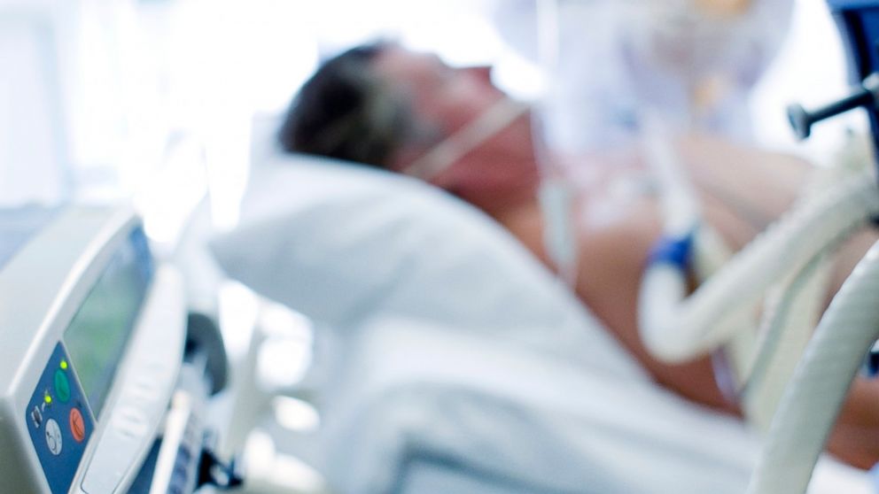


From ABC News


The first patient to communicate with Owen was a 26-year-old patient named Kate Bainbridge. In 1997, a deadly viral infection caused Bainbridge to go into a coma. Using positron-emission topography (PET) to examine a brain area called fusiform face area (FFA), which is activated whenever one sees a familiar face, Owen decided to investigate what would happen to Bainbridge if she saw the face of someone she knew. Bainbridge showed significant brain function according to the neuroimaging data. By 2010, she was in a wheelchair, but otherwise active following rehabilitation. Despite similar responses in other patients in comas, he and his research has still been met with backlash (Cyranoski, 2012). Both scientists and the general public have shown dislike for Owen’s brain imaging on patients in a vegetative state. Owen's method raises many questions for physicians, for it radically changes the standard procedures of the treatment of patients in comas. One question it raises is what type of effect will his research have on a family's or clinician's decision to end a life. “If a patient answers questions and demonstrates some form of consciousness, he or she moves from the 'possibly allowed to die' category to the 'not generally allowed to die' category,” says Owens. This will put the general public in an awkward position when it comes to processing the death of a loved one. Some will be given hope, whereas others will be burdened financially by the prospect of keeping them alive for these not particularly understood signs exhibited by the patient. Also, this leads to the major concern: How much say does the patient have in their death.


Owen is now planning on using an electroencephalogram (EEG) to complement these imaging studies. EEGs also have the advantage of being portable, cost-effective and relatively fast. Owen will examine 25 people a year to prove that this may be a way to communicate with patients in vegetative states. However, one must ask oneself one question: Is this enough to say that what is being seen in a brain image or EEG is an accurate representation of a patient’s desires and needs? Could it possibly be simply an automatic versus intentional response?


Caption: fMRI images comparing a patient given the diagnosis of existing in a vegetative state to healthy volunteers. All participants in the study completed two imagery tasks under the direction of Adrian Owen. From Owen et al


Determining death based on brain death is flawed according to both sides of the debate. There is no reliable universal way to determine brain death unless the entire brain has been destroyed and the circulatory and respiratory systems have also stopped functioning. Moreover, people pronounced brain dead can “survive” on life support for extended periods of time. This is because the “survival” of the body is not solely dependent on brain functions; it’s also dependent on circulatory and respiratory functions, which can exist even if the brain is not functioning at all. But is living without brain function, or more importantly a cognitive life, living? As of the present, one year later, Jahi McMath is still surviving with the help of a ventilator. There is even recent videotape footage of her moving her foot and hand at the request of her mother. So is this girl who was declared neurologically dead really dead? Doctor say “yes,” her parents say “no,” and I say the uncertainty surrounding her case makes it better to be safe than sorry to keep her on the ventilator. According to Georgia Law, Statute 31-10-16, death is either irreversible cessation of circulatory and respiratory functions or irreversible cessation of all functions of the brain, including the brain stem. Advanced directives will be recognized first and foremost, for Georgia replaced its Living Will and Durable Power of Attorney for Health Care as of July 1st, 2007. Therefore, if a patient were to write both a living will and advanced directive of contradictory orders for what to do in the face of their impending death, the advanced directives will be followed. (These laws can be seen here and here). However, if a person were to undergo Owen’s test and brain activity were to be found, an advanced directive, in the state of Georgia would not be able to be honored. The legal consequences of utilizing such a technology complicate things even further. Furthermore, with all these varying opinions and stakeholders in Jahi McMath’s death declaration, this prompts the question: Who has the right to be involved in this kind of decision anyways?


Reference


Owen, A. M.; Coleman, M. R.; Boly, M.; Davis, M. H.; Laureys, S.; Pickard, J. D. Detecting Awareness in the Vegetative State. Science 2006, 313 (5792), 1402.


Caplan, Arthur. "Brain Death Really Is Death." Time. Time, 3 Jan. 2014. Web. 25 Feb. 2015.


Cyranoski, David. "Neuroscience: The Mind Reader." Nature.com. Nature Publishing Group, 13 June 2012. Web. 06 Mar. 2015.


"Organ Donation/ Transplantation." Education Organ Donation / Transplantation End of Life Care: When Is a Person Really Dead? American Life League, n.d. Web. 25 Feb. 2015.


Pernick, M., 1999, “Brain Death in a Cultural Context: The Reconstruction of Death, 1967–1981,” in in S. Youngner, R. Arnold, and R. Shapiro (eds.), The Definition of Death: Contemporary Controversies, Baltimore, MD: Johns Hopkins University Press: 3–33.


Vincent, C. (2012). The essentials of patient safety. Download disponibile all’indirizzo www1. imperial. Ac. uk/medicine/about/institutes/patientsafetyservicequality.


Want to cite this post?


Green, A. (2015). Is Brain Dead Really Dead? The Neuroethics Blog. Retrieved on , from http://www.theneuroethicsblog.com/2015/03/is-brain-dead-really-dead.html

Tuesday, December 4, 2012

The Future of Intelligence Testing


Few people I know actually enjoy standardized tests. Wouldn’t it be great if technology could eliminate the need for bubble-in forms and Scantron sheets? How nice would it be to simply go in and get a snapshot of your brain to find out how smart you are? Imagine walking into the test center, signing on the dotted line, getting a quick scan, and walking out with your scores in hand, helping you gain admittance into a college or land your next job. No brain-racking questions, no tricky analogies, and no obscure vocabulary. Goodbye SAT, hello functional magnetic resonance imaging (fMRI).


Image from 


http://theturingcentenary.files.wordpress.com/2012/06/brain-functions.jpg    


In the general, there have been two types of intelligence studies: psychometric and biological. Biological approaches make use of neuroimaging techniques and examine brain function. Psychometrics focuses on mental abilities (think IQ tests). Dr. Ian Deary and associates suggest that a greater overlap of these techniques will reveal new findings. In their paper, 'Testing versus understanding human intelligence,' they state:


“The lack of overlap between these approaches means that it is unclear what the scores of intelligence tests mean in terms of fundamental biological processes of the brain.”[2]


Applying psychometric analysis techniques (IQ tests) coupled with advanced imaging has the potential to reveal the locations of “higher” cognition and neural processing. I expect that future technologies will have incredibly improved resolution, which will allow scientists to see not only what regions the brain uses but also the exact pathways that are activated for each action. By understanding which pathways are utilized for specific tasks, it may be possible to identify which genes as well as environmental factors (nutrition, education) are responsible for their development. This can lead to programs dedicated to training specific areas of the brain (several of which already exist) [5], and perhaps even drugs that foster development.


I believe it is increasingly important to consider the implications of a technology powerful enough to quickly evaluate an individual’s level of intelligence. However, before I get there, it is necessary to first explain what we can and cannot currently do. What we cannot currently do is use neuroimaging to determine how smart you are. What we can currently do is use neuroimaging to see what parts of your brain contribute to how you process information, access memories and function [3].


Localizing Intelligence


Different parts of your brain do different things and none of these brain regions work in isolation. Some regions contribute to eating, seeing, and other regions play a role in “intelligence”. The varying techniques of imaging-based testing search for different correlates of intelligence [4] (i.e., general intelligence, problem solving, learning abilities). Developments in imaging technologies have improved our ability for greater analysis, allowing for the study of both damaged and healthy brains. For example, MRI studies have found that the volume of gray matter correlates to intelligence, providing evidence for generalizations made regarding brain volume and intelligence [7]. A 2006 study of 100 postmortem brains examined the relationship between an individual’s Full Scale Wechsler Adult Intelligence Scale (WAIS) score and the volume of their brain regions. The factors they considered important to the relationship between brain size and intelligence were age, sex and hemispheric functional lateralization (They found that general verbal ability was correlated with cerebral volume in women and right-handed men. They did not find a relationship between ability and volume in with every group, however).


Additionally, PET and fMRI studies have revealed more information regarding the functionality of certain regions of the brain. By recording and interpreting the brain activity of subjects as they complete a variety of tasks, researchers are able to draw inferences based on the performance in the types of task (and thus, the type of intelligence) that calls on particular areas of the brain.  This is interesting, as knowing how parts of the brain are utilized may reveal more information about the structure and hierarchy used in neural development. It also may provide interesting information regarding the pathways of neural signals throughout the nervous system. Image-based testing may allow researchers to discover why certain neurons are connected, if they are indeed aligned in a purposeful manner and consequently, how to repair such pathways when they are damaged.


Image from http://news.wustl.edu/news/Pages/24068.aspx

A study from Washington University in St. Louis has shed light on how our brains utilize various networks for performance with working memory tasks [1]. They described a mechanism, global connectivity, which coordinates control of other networks. In a sense, global connectivity is the CEO of your brain, insuring that all components of your system are functioning and allowing for effective control of thought and behavior. Specifically, they found that a region of the lateral prefrontal cortex (LPFC), whose activity has been found to predict working memory performance, employs global connectivity. They report that,


“critically, global connectivity in this LPFC region, involving connections both within and outside the frontoparietal network, showed a highly selective relationship with individual differences in fluid intelligence. These findings suggest LPFC is a global hub with a brainwide influence that facilitates the ability to implement control processes central to human intelligence.”


This fascinating study identified a very specific characteristic of the brain (the global connectivity of the left LPFC) that suggested investigators were accurately able to predict fluid intelligence (where fluid intelligence refers to reasoning and novel problem solving ability) [4].


Potential Issues


We are learning more about the brain and the biological bases for intelligence every day. We have expanded our understanding of memory, cognitive thought and neural computation [2,6]. Our understanding of imaging techniques and what we can learn from them continues to grow. It may very well be possible to someday use neuroimaging to evaluate an individual’s intelligence. It is becoming more widely accepted that a neurobiological basis for intelligence exists (at least for reasoning and problem-solving) [4]. At the same time, the success of these intelligence studies presents ethical issues. Gray et al. pose the question, “Is it ever ethical to assess population-group (racial or ethnic) differences in intelligence?” While little variation has been found between racial groups, the public perception of intelligence studies has been negatively impacted by concerns of racism [4]. It is important to consider the consequences of studies that investigate intelligence differences in population-groups (racial, ethnic, and socioeconomic status). Gray states that it is not necessary to consider race when exploring the neurobiological bases of intelligence. The majority of variation occurs within a racial group and not between them. However, if a study were to investigate race and intelligence, Gray states that it will be necessary to have consent as well as active support from the target groups (i.e., financial support).


There are in fact studies that have investigated test score differences associated with race. Claude Steele, Ph.D., a professor of social psychology at Stanford University, discussed the test performance differences of white and black Americans. In his interview with PBS, Dr. Steele explained that a serious gap in test scores exists between whites and blacks, citing 100-point differences on the verbal and quantitative sections of the SAT. This is a concern for policy makers who are responsible for maintaining a standard level of education, as these low scores may help identify areas that need improvement. However, the negative effect of these findings is due to something Dr. Steele refers to as “stereotype threat.” He explains that when a salient negative stereotype about a group that you identify with may apply (i.e., lower SAT scores), when you are in that test situation the prospect of matching the stereotype can be distracting and upsetting. This stereotype threat impacts test taking, undermining test performance.


When considering the neurobiological bases of intelligence, it may be harder to escape these stereotypes. It may someday become known which genes code for higher intelligence, and thus higher test scores. Already, we have begun studying what regions of the brain relate to intelligence test performance. The next step will be discovering which genes code for the development of those regions, then connecting the dots from genes to intelligence. Furthermore, an understanding of the environmental and social influence that control the activation of these genes (and thus development) will be needed. Future generations may have stereotype threats that are based on genes rather than groups. When you know that you carry the genes for a certain level of intelligence, you may find yourself doubtful of your ability to perform above the level predicted by your genome and fall short of your potential.


There is a lot to be learned regarding how our genes relate to intelligence and by understanding how different gene pools code for their “smarts,” our understanding could grow immensely. However, as I mentioned earlier, suggesting that one group is genetically hard-coded to be smarter than another will have enormous implications. Science and research are going to continue pushing this ethical boundary and I predict that we eventually will be able to find many links between genes and intelligence. It is both beneficial and crucial to discuss how genes and intelligence should be studied now, before the research takes place. We have a unique opportunity where ethics can lay the guidelines before this technology emerges.


There are exciting possibilities that neuroimaging intelligence tests may bring. On the one hand, we can learn so much about the brain, how we think and how we can make it better. On the other hand, it may drive ethnic and racial groups, as well as socio-economic groups, farther apart. So we really have to ask ourselves, is that the price we have to pay to not fill out another Scantron sheet?


Want to Cite This Post?

Craig, E. The Future of Intelligence Testing. The Neuroethics Blog. Retrieved on from, http://www.theneuroethicsblog.com/2012/12/the-future-of-intelligence-testing.html


Related articles


http://headblitz.com/what-brain-scans-might-replace-iq-tests-in-the-future/

http://www.mobiledia.com/news/142925.html

http://www.medicaldaily.com/articles/11216/20120801/intelligence-mri-iq-test-brain.htm

http://www.psychologytoday.com/blog/finding-the-next-einstein/201202/could-brain-imaging-replace-the-sat

http://en.wikipedia.org/wiki/Neuroimaging_intelligence_testing


References


1. Cole, M. W., Yarkoni, T., Repovs, G., Anticevic, A., & Braver, T. S. (2012). Global connectivity of prefrontal cortex predicts cognitive control and intelligence. The Journal of neuroscience : the official journal of the Society for Neuroscience, 32(26), 8988–99. doi:10.1523/JNEUROSCI.0536-12.2012

2. Deary, I. J., & Caryl, P. G. (1997). Neuroscience and human intelligence differences. Trends in neurosciences, 20(8), 365–71. Retrieved from http://www.ncbi.nlm.nih.gov/pubmed/9246731

3. Duncan, J. (2000). A Neural Basis for General Intelligence. Science, 289(5478), 457–460. doi:10.1126/science.289.5478.457

4. Gray, J. R., & Thompson, P. M. (2004). Neurobiology of intelligence: science and ethics. Nature reviews. Neuroscience, 5(6), 471–82. doi:10.1038/nrn1405

5. Hackman, D. a, Farah, M. J., & Meaney, M. J. (2010). Socioeconomic status and the brain: mechanistic insights from human and animal research. Nature reviews. Neuroscience, 11(9), 651–9. doi:10.1038/nrn2897

6. Prabhakaran, V., Rypma, B., & Gabrieli, J. D. E. (2001). Neural substrates of mathematical reasoning: A functional magnetic resonance imaging study of neocortical activation during performance of the necessary arithmetic operations test. Neuropsychology, 15(1), 115–127. doi:10.1037//0894-4105.15.1.115

7. Witelson, S. F., Beresh, H., & Kigar, D. L. (2006). Intelligence and brain size in 100 postmortem brains: sex, lateralization and age factors. Brain : a journal of neurology, 129(Pt 2), 386–98. doi:10.1093/brain/awh69