Pages

Tuesday, March 26, 2013

Hubris and Hope for Engineering Brains

"Living organisms are nothing more than complex biochemical machines." [1]


The above statement, or at least the thought, is something that gets thrown around in biology and especially bioengineering. And it's a very empowering thought: the living thing in front of me is governed by the same physical laws that govern the rest of the universe, including the machines we build.  It doesn't have some sort of supernatural vital force flowing through it, just fats and proteins and DNA and small molecules.  We can use this.  We can fix ourselves when we are sick, we can design new life forms to do our bidding.  It's all very exciting.


We might even go as far as to engineering a brain.  The task would be difficult, but the reward tremendous.  Brains are very good at performing difficult computations that top of the line AI is struggling with, and biological neurons tend to use less resources than their artificial cousins. Building a wet, squishy, thinking machine, designed to perform one specific task and to perform that task very well, would be a great boon to autonomous robots, power grid management, and thousands of other applications. Hey, we've already engineered neural tissue to be part of an art project.


Ionat Zurr and Oron Catts of Symbiotica
However, Ionat Zurr and Oron Catts of the SymbioticA biological art center in Perth, Australia, are quick to bring up the fact that thinking of living systems as machines brings with it a lot of additional baggage. [2] Machines don't get a lot of respect. They are 'tools', 'playthings', to be used without reverence or regard to their own desires or wants.  What could a hammer possibly want?  If somehow it did want something, why should we care?  What could we do to an electrical circuit that could make us feel guilty?  By extension, why should we care about the wants of individual cells, or organs, or even living organisms?


One critique (of many possible critiques) of this mechanical view of neural systems is that it supposes an impossible level of understanding.  This isn't an attack on determinism or naturalism, so much as an acknowledgement of how much we don't currently know, and how much we are likely to know in the near future.  It is simple enough to understand a hammer to the point that it can be used effectively - you physically move it as an extension of your hand, and the metal end drives in a nail.  A sled dog, on the other hand, requires an immense understanding to 'use' properly, so that the dog acts as an extension of your own will. [3] The argument follows that we should be prepared to think of biological systems, and neural systems especially, as being too difficult to engineer in their entirety.  Without this perfect knowledge, how can we be sure that we haven't created a slave that can't even express its own suffering?


Tools can be adorable in addition to being functional.  From here
This living complexity and danger of moral atrocity is most severe in neural systems.   Much like all biological systems, there is a lot about the behavior of neural systems that remains unknown.  But even the 'knowns' in neural systems seem to act to prevent understanding.  Neural systems interconnect directly with every aspect of the body, and the environment beyond the body, so the effective environment that neural systems respond to (and thus, the number of variables engineers need to account for) is especially vast.  Additionally, neural systems are famously malleable - they adapt themselves to meet the demands of their environment, meaning that even if a neural system is understood to some level of satisfaction at one point in time, it could very well surprise you later on. This, combined with the general scientific and cultural notion that neural systems are responsible for such ethically nasty things as pain and suffering, makes it very difficult to be sure something terrible won't happen when we start to design with neural tissue.


However, the discussion of what is unknown, or perhaps (practically) unknowable, about biology and neural systems should also include mention of the techniques that engineering has created in order to deal with overwhelming complexity.  For example, modern software engineering projects are so large that is impossible for any one person to understand the entire project all at once. Instead, the insurmountably large project is broken down into individual components or objects, which in turn are built of smaller and smaller objects. The idea is to test the system at each level of description, so that unexpected behaviors can be identified at their origin and fixed.


The fly olfactory sysem- from [4].  
The beginnings of this compartmentalization can even be seen in neuroscience work.  A fantastic example is Professor Larry Abbott's work to provide functional descriptions of different levels of the fly olfactory system [4], where three successive layers of connections were described as three different functional blocks (noise reduction, normalization, and a reservoir computing style state expansion). While there wasn't any discussion on what processes are necessary to maintain those functions in the face of unexpected interactions with other systems, or plastic changes in the blocks themselves (not to mention that this occurred in the humble fruit fly), the modular architecture used implies that such techniques could be used to engineer neural systems to behave in similar ways. So perhaps the engineers are up to the challenge of harnessing the computational capabilities of living tissue, in a way that is robust against the construction of entities that we would look upon with pity.


-------------

References

[1] Note that this quote refers to how biologists, not the author, views living organisms http://informatics.indiana.edu/larryy/al4ai/papers/Langton.ALife.pdf


[2] Catts, Oron, and Ionat Zurr. "The Vitality of Matter and the Instrumentalisation of Life." Architectural Design 83.1 (2013): 70-75.


[3] The mechanical view of life can be made more practical and perhaps even less cold by making sure to include things like "exceeding the physical limits of a tool degrades performance" and "suffering decreases efficiency."  The ideal living tool wouldn't suffer from it's use, but would enjoy pleasing it's user- or perhaps master is a better word here-  and seek to improve it's own performance.  Of course this could easily horrify someone with the imagery of a slave that participates in it's own slavery, unable to even perceive what a better life might be.


[4] Keene, Alex C., and Scott Waddell. "Drosophila olfactory memory: single genes to complex neural circuits." Nature Reviews Neuroscience 8.5 (2007): 341-354.


[5] This work includes the paper "Generating sparse and selective third-order responses

in the olfactory system of the fly", as well as work that is currently under review.  Dr. Abbott gives a fantastic talk.


Want to cite this post?

Zeller-Townson, RT. (2013). Hubris and Hope for Engineering Brains. The Neuroethics Blog. Retrieved on from http://www.theneuroethicsblog.com/2013/03/hubris-and-hope-for-engineering-brains.html

Tuesday, March 19, 2013

Dare to be different: Defense of the research of sex differences

By Guest Contributor, Emory Neuroscience and Animal Behavior Graduate Student Katy Renfro


In a recent article published in the journal Neuroethics, Dr. Rebecca Jordan-Young and Dr. Raffaella Rumiati argue that current research on sex differences is “both unscientific and far from politically neutral,” and should be abandoned. [6] This article reflects many of the current conversations on the ethical implications of researching sex differences, which have largely focused on how results of these studies can be misappropriated to support sexist agendas. I cannot argue against the legitimacy of these concerns, and as researchers, we must always be careful to present our findings in a balanced and accurate manner so as to better combat misinterpretations and misrepresentations of data. However, we must also keep in mind that just as science has the potential to influence social and political conversations, this is a bidirectional relationship, and politics also have the power to misinform science.


In their paper, Jordan-Young and Rumiati argue that current research on

sex differences is "both unscientific and far from politically neutral." [6]


In their article, Dr. Jordan-Young and Dr. Rumiati offer two alternatives to sex difference research; the avenue they appear to more actively promote is to ignore sex differences altogether, in favor of researching differences between groups of people, such as socioeconomic or occupational classes. I believe this suggestion is an example of political motivations influencing science. Due to past and current sex/gender inequalities, there continues to be a strong push for leveling the sex/gender playing field. Although I am (of course) a strong supporter of equality between the sexes, I think it is misguided to push for the abandonment of a fruitful field of research because of the possible political implications of its results; indeed, I find the suggestion that we should halt inquiry in this area both unethical and unscientific.


Below, I provide rationale for why abandoning the study of sex differences would have ramifications for basic science research, and I then offer ways in which we as scientists can better address the potential political implications of the results of our studies.


Brain, body, and behavior


In Dr. Jordan-Young and Dr. Rumiati’s article, the researchers distinguish the body from the brain, asserting that although there are clear phenotypic differences between men and women that are due to hormonal effects, these differences are not representative of neural or behavioral differences. They support this claim by setting up a thought experiment: if a group of men and women were given images of male and female genitalia and were asked to categorize the images by sex, they would easily be able to do so; however, the same would not hold true for the brain—there are no gross, morphological indicators of the prototypical “male” and “female” human brain.


A different thought experiment: would it be possible to categorize men and

women's gastrointestinal tissues by sex?

In response, I will propose another thought experiment. If one were to gather a group of men and women, and provide them with photographs of men and women’s gastrointestinal (GI) tissues (i.e. their stomachs), would they be able to categorize them by sex? I am fairly sure the group would perform this task at no better than chance. However, if one were to use modern scientific techniques to assess the pH and enzymatic profiles of these stomachs, and examine these profiles with help from the literature, it would become immediately apparent which belonged to the men and which belonged to the women, as men and women differ in acidity of gastric secretions and in activity of a number of gastric genes. [8,5] In fact, if one were to assess activity of even just one enzyme: alcohol dehydrogenase, he/she would most likely be able to differentiate the GI tissues by sex, as this enzyme is much more active in men than women. If we take this a step further, and look for an alternate macro reflection of this difference that is not evident in the organ’s physical appearance, we could ask whether differences in this gene are related to behavioral sex differences, and the answer would be yes. When men and women drink comparable amounts of alcohol, men (due to greater enzymatic activity) have significantly lower blood alcohol content (BAC), and thus can drink markedly greater amounts before experiencing the adverse side effects of high alcohol consumption. [4, 7, 2, 3]


Because researchers looked deeper than the organ’s physical appearance and revealed these sex differences, they then had clearer directions to follow when looking for the mechanisms underlying these differences, as a sex difference indicated that prenatal and/or circulating hormones could be at play. Indeed, it has been found that altering hormone levels affects expression of alcohol dehydrogenase (e.g. [1]). There are, of course, also environmental and other biological factors that modulate these sex differences in enzymatic activity and alcohol consumption (e.g. overall body size, regularity of alcohol consumption); however, recognizing that there is a sex difference allows for more careful investigation into underlying mechanisms, as one can use this information to generate hypotheses and design experiments that specifically examine the role of hormones. To ignore the sex difference would be to ignore an integral piece of the scientific picture.


There are reported sex differences in the prevalence of nearly every major mental illness. [4]


Our internal organs cannot be completely divorced from our phenotypes; our body cannot be completely divorced from our brain, and both the brain and the body affect and are affected by our behavior. Just as recognition of sex differences provided a window into the mechanisms underlying alcohol consumption and alcohol-related disease prevalence, so it can provide us with clear avenues through which to explore the neural mechanisms of behavior. For example, there are reported sex differences in the prevalence of nearly every major mental illness [4]; ethically and scientifically, we cannot afford to deny these differences if we seek to better understand the basic biological mechanisms of illnesses such as depression, substance use disorders, and generalized anxiety disorder. Indeed, only after we understand the biological mechanisms of a disease can we better develop novel and effective treatments for it.


Conclusion and future directions


Knowledge always carries with it the potential for misuse; this is true for all scientific advancements, whether they be technological, chemical, or biological in nature. In using the example of these discoveries with the stomach, one could imagine that these results could be used to support differential regulations on alcohol consumption between the sexes, or to portray woman as “too pure” to consume alcohol “like a man.” However, it is these threats to equality that we should specifically work to eliminate—not the science of sex differences.  The discovery of “difference” does not have to mean the discovery of ammunition to fuel societal inequalities. Indeed, difference itself carries with it no moral value or attribution of goodness/badness; it is culture that assigns value to these differences. Some of the strongest resources we have to combat misrepresentations of data are social media sources. As researchers, we should take on the responsibility of writing accurate and politically neutral pieces on our research so the public is presented with a clear picture of what we did in the experiment, what was found, and what it means. Through social media avenues, such as youtube videos, blogposts, or twitter links, we can promote these pieces and reach larger audiences. It should also be our responsibility to monitor the way our results are interpreted by outside sources and to counter misrepresentations—this is a responsibility that some researchers have already taken on (for example, see Dr. Lisa Diamond’s defense of her research on sexual fluidity against the National Association for Research and Therapy of Homosexuality’s misuse). In an increasingly “flattened” world, knowledge spreads within minutes; our job as scientists should not be to stop learning or to stop the dissemination of knowledge, but rather to make sure that we are among those disseminating it, so that it can be done so in a responsible manner.


References

[1] Aasmoe, L, & Aarbakke, J. (1999). Sex-dependent induction of alcohol dehydrogenase activity in rats. Biochemical Pharmacology, 57(9), 1067-1072.


[2] Baraona, E, Abittan, C S, Dohmen, K, et al. (2001). Gender differences in pharmacokinetics of alcohol. Alcoholism: Clinical and Experimental Research, 25(4), 502-507.


[3] Ceylan-Isik, A.F., McBride, S.M., & Ren, J. (2010). Sex differences in alcoholism: who is at a greater risk for development of alcoholic complication? Life Sciences, 87, 133-138.


[4] Eaton, N. R., Keyes, K. M., Krueger, R. F., et al. (2012). An invariant dimensional liability model of gender differences in mental disorder prevalence: Evidence from a national sample. Journal of abnormal psychology, 121(1), 282-288.


[4] Frezza, M, di Padova, C, Pozzato, G, et al. (1990). High blood alcohol levels in women. the role of decreased gastric alcohol dehydrogenase activity and first-pass metabolism. The New England Journal of Medicine, 322(2), 95-99.


[5] Gandhi, M., Aweeka, F., Greenblatt, R.M., Blaschke, T.F. (2004). Sex differences in pharmacokinetics and pharmacodynamics. Annual Review of Pharmacology and Toxicology, 44, 499-523.


[6] Jordan-Young, R. & Rumiati, R.I. (2012). Hardwired for sexism? Approaches to sex/gender in neuroscience. Neuroethics, 5(3), 305-315.


[7] National Institute on Alcohol Abuse and Alcoholism (1997). Factors influencing alcohol absorption and

metabolism. Retrieved from: http://pubs.niaaa.nih.gov/publications/aa35.htm


[8] Whitley, H., & Lindsey, W. (2009). Sex-based differences in drug activity. American Family Physician, 80(11), 1254-1258.


Want to Cite this Post? 

Renfro, K. (2013). Dare to be different: Defense of the research of sex differences. The Neuroethics Blog. Retrieved on Monday, Day, Year, from 

Monday, March 11, 2013

Neuroethics Journal Club: Imaging Pedophilia

The form of argument is familiar: X is bad, very bad. We can maybe stop X, but in order to do so, we’ll have to compromise our values just slightly. We’re not happy about it, but after all, X is very, very bad.


At this month’s Neuroethics Journal Club led by psychology graduate student Katy Renfro, X was pedophilia, and the topic of discussion was a new technique to decrease pedophilic sexual desire through fMRI-based brain computer interface (BCI). The title of the paper –Renaud et. al’s “Real-time functional magnetic imaging—brain–computer interface and virtual reality: promising tools for the treatment of pedophilia” – immediately raised two questions in my mind (which, as I eventually realized, were probably not the right ones). First, is it going to be possible to Clockwork Orange pedophiles? Second, if so, should it be done?


A treatment for pedophilia?


So What Did They Do?

Nothing, actually. As one participant put it: “this is basically the beginning of a fishing expedition.” This particular fishing expedition was for a new, fMRI-integrated version of an old technique – biofeedback – in which a patient receives physiological information in order to help control subtle thoughts or behaviors. For instance, a person suffering from anxiety might practice relaxation techniques while having their heart rate and blood pressure measured. This physiological feedback can help indicate which techniques are most successful, helping the patient to better manage their anxiety.


In the context of pedophilia, Renaud et. al suggest a biofeedback technique premised on activation of the anterior cingulate cortex (ACC). The ACC, the authors argue, is associated with impulse control and sexual arousal. If pedophiles could learn to decrease activation in the ACC, then, it follows that sexual attraction to children (or at least the impulse to act on that attraction) might be diminished.


So how to decrease ACC activation? The authors propose placing pedophiles in an fMRI (voluntarily, of course), showing them computer-generated images of children (the “virtual reality” from the paper’s title), and mapping the children’s movements onto ACC activation. So as ACC activation decreases, a computerized child might raise its hand, and the pedophile would know that they had successfully decreased ACC activity. To help things along a bit, the authors also suggest a “strategy of covert mental rehearsal,” meaning the patient continually thinks about the terrible things that would happen to them if they were caught acting on their desires.


The theory behind biofeedback.

Does It Work?

I should reiterate that Renaud et. al didn’t actually do any of this: they just proposed the methodology. On the fMRI biofeedback fishing expedition, they haven’t even gotten around to buying the fishing poles. But there’s enough detail in their methodology that it’s possible to guess at the likelihood that it would succeed, something that the discussion participants were happy to do.


Concerns fell into a few categories. First, there was widespread agreement that ACC activation is too simplistic to be a reliable measure of sexual desire. Not only is the neurobiology of sexual desire poorly understood in general, but the ACC is involved in many non-sexual functions including error detection, empathy, and suppressing undesirable thoughts, functions that may not be desirable targets of biofeedback. Second, attempts to modify sexual desire, most notably homosexual desire, have historically been unsuccessful. One participant noted the difficulty this poses for fMRI biofeedback: either pedophilic sexual desire is basically similar to other forms of human sexual desire, in which case any form of “conversation therapy” should be expected to fail; or, pedophilic sexual desire is fundamentally different from other sexual desire, in which case there is no reason to target regions like the ACC that have been implicated primarily in studies with non-pedophiles. Finally, participants raised concerns regarding ecological validity. A man in an fMRI scanner, looking at computerized images of children whose movements he partially controls with his brain, wearing a cylinder around his penis to measure arousal and a camera tracking his eye movements, in a room with psychiatrists and neuroscientists and fMRI technicians, is not the same as a real-life encounter with a child. There is reasonable concern, then, that fMRI biofeedback for pedophilia would not be effective in the real world even if data showed it to be effective in the laboratory.


Perhaps not the same as viewing an emu in the Australian wilderness.

But What If It Did Work?

Well, it still might. It’s probably unfair to criticize a person’s fishing technique before they’ve even gotten to the lake, and Renaud et. al’s paper is a proof-of-concept proposal, not a meticulously worked out solution. Several participants suggested that fMRI biofeedback is worth pursuing, even if it only starts paying material dividends five or ten years down the line. So it’s worth asking: if such a technique could be used successfully with pedophiles, is there any reason why we should be concerned about implementing it?


fMRI biofeedback is not Clockwork Orange: nobody’s eyes are forcibly pried open, nobody is socialized through the infliction of horrific pain. The sense in which it’s like Clockwork Orange, and in which it might make some people uncomfortable, is that it uses technology to change behavior. There’s nothing new about this – all drugs, all therapies set out to change behavior – but there are plenty of reasons why super-effective behavioral modification techniques are scary. Luckily, fMRI biofeedback doesn’t seem to meet that criterion, so there’s probably little risk of slipping into a behavioral-control dystopia.


A more relevant concern may be the implications of successful fMRI biofeedback on how the law understands and punishes sex offenses against children. In a previous Neuroethics Blog post, Cyd Cipolla has argued that the perceived special heinousness of sex crimes against children, along with pedophilia’s incurability by any current therapy, has resulted in a set of laws premised on the notion that pedophiles “are not on the extreme edge of criminal behavior we imagine them to be, but are, in fact, in a category of their own.” These laws include provisions for lifetime surveillance and community notification following certain types of sex offenses, as well as the possibility of indefinite civil commitment for some offenders. According to Cipolla, the real issue at stake is not criminal responsibility – which is a given – but empathy: “How does knowing about the cause… help us to move towards real empathetic solutions even while maintaining guilt?”


There are still reasons for caution, however. To paraphrase one participant: we need to reflect on whether our goal is really create effective therapies, or if we’re ultimately more interested in “making ourselves feel better.” Therapy for pedophiles is enormously attractive in that it allows us to tell ourselves we’re doing something about a behavior that, for most of us, is horrifying. But to feel like we’re doing something about pedophilia is not the same as actually dealing with it – and especially in politics, the two can easily become conflated. The barriers to fMRI biofeedback are significant: we don’t know that much about sexual desire, we don’t know that much about pedophilic sexual desire in particular, there are plenty of questions about fMRI reliability, and fMRI feedback in particular is a totally new and untested technique. There are no clear answers, but plenty of questions; and given the sensitivity of this particular issue, there is a danger that limited empirical results could be blown up into significant political pressure to implement fMRI biofeedback on a large scale. That would be a shame, if for no other reason than it would discourage us from asking difficult questions about how we can most effectively deal with sexual violence.

Life in Death: The Neurobiology of Near-Death Experience


Surely you’ve seen this in film or read something like it in fiction. The victim of a tragic accident is critically wounded and rendered unconscious, let’s say in a motor vehicle crash. Within minutes, an emergency medical team arrives at the scene and drags his limp frame out of the crumpled car. Paramedics surround him, now splayed out the roadside, working frantically to restore his breathing and pulse. Each passing second is precious. With every tick of the clock, his vital organs lose more of their precious oxygen and energy required to function. As the victim’s brain becomes increasingly hypoxic (lacking oxygen), his neurons execute coordinated self-destruct programs, and the chance of restoring his consciousness diminishes. Lacking a heartbeat, respiration, and autonomic reflexes that indicate brainstem function, he meets criteria for clinical death (CD). The medical team continues to toil, and the hope of recovery dims for our fallen protagonist.


Here’s where the story gets predictably paranormal and cinematic. I’ll offer three pop depictions of the dying process, but I’m sure the reader can let her imagination run wild and come up with many others. Option 1: At this point in the scene, a wispy duplicate of the victim separates off and ascends from his physical body, floating a few feet above. The deceased’s perspective is like a detached observer of the physical world. Our hero watches his lifeless body with equanimous curiosity while the medical team down below continues their attempts at resuscitation... Option 2: A slight variation on this would be to have the deceased completely exit normal reality and enter an otherworldly realm of light, visions, and divine beings... Option 3: A montage of past life events ensues, his most egregious failures revisited and relived, decades of experience compressed into minutes. Finally, the vignette concludes when the patient is revived and his disembodied consciousness snaps back into his body.


These scenarios are so consistent and so prevalent in our culture that we may overlook their commonality as nothing more than the over-played makings of Hollywood. But intriguingly, many real-world survivors of CD report strikingly similar near-death encounters (NDEs) and out-of-body experiences (OBEs) [1] to those synthesized for our viewing pleasure by popular culture. In fact, civilizations and religions throughout history share remarkably common eschatology (theories of death and the soul), despite huge differences between other aspects of their culture and dogma. Ancient Egyptians and Tibetan Buddhists, for example, both had funerary texts describing the proximal process of death in which one’s consciousness exits the body, translated as the “Book of the Dead,” or the “Bardo Thodol,” in Tibetan. Fifteenth century Dutch painter, Hieronymus Bosch, depicts the soul ascending toward a tunnel of light in his famous work, Ascent of the Blessed. All three Abrahamic religions believe that the soul departs from the physical body and transits to a divine (or not-so-divine) world. It seems unlikely that these commonalities originated from a single ancestral source, memes propagated across many miles and many millennia. Instead, they may have resulted from parallel evolution, in which individuals from each culture recounted their near-death experiences, which were then reinterpreted to fit neatly into their own cultural milieu. Here, in the 21st century, we are undergoing the same parallelism; the difference being that medicine and neuroscience are our secular framework into which NDEs must fit.


"Ascent of the Blessed" by Heironymous Bosch; retrieved from Wikimedia Commons


As I alluded to above, CD requires three things: 1) lack of heartbeat; 2) absence of respiration; and 3) no brainstem activity, typically assessed by the pupillary light reflex. In recent years, improvements in modern resuscitation technologies, such as automated chest compressors, have resulted in many more CD survivors today than only fifty years ago. As a result, studies over the past few decades have been able to gather lots of anecdotal information from cardiac arrest survivors about the nature of their experiences (if indeed they can recall them) during the time in which they were pronounced clinically dead. American psychiatrist, Dr. Raymond Moody, was the first to document such cases in the late 1970’s [1]. Among 150 critically ill patients with NDEs, he found that survivors typically reported feelings of peace, seeing a tunnel and a bright light, as well as separation from the body. These stories bear a striking resemblance to those described in ancient cultures and world religions.


The first prospective study to report on cognitive processes during cardiac arrest found similar anecdotes in survivors [2]. In the most exhaustive study published to date (344 subjects), its authors report that around 12% of all cardiac arrest survivors had NDEs [3]. Others suggest that as many as 20% or more report cognitive processes and memories from the period of resuscitation [4]. In addition, survivors with NDEs report long-term improvements in social functioning and psychological well-being 2 years post-recovery compared to non-NDE cardiac arrest survivors [2]. Perhaps the most intriguing anecdotes of all come from cardiac arrest survivors who report OBEs similar in nature to our fictional story from earlier. In the study by van Lommel, et al., 24% of patients with NDEs also reported being able to watch and recall events from their resuscitation. In one such anecdote, the patient was actually able to identify the nurse who had removed his dentures during emergency revival efforts and correctly specify where she had placed them. Many survivors and medical professionals tell of similar experiences, and it is likely that such cases are underreported due to fear of stigmatization.


So, now that we’ve established that NDEs and OBEs are more prevalent in the 21st century than any other time in human history, the quest for a neurobiological explanation begins. There are many theories out there that seek to justify the clear phenomenology of these experiences, including but not limited to 1) tunnel vision, 2) disembodiment, 3) hallucinations, 4) feelings of profound serenity, and 5) life history recollection. Before delving into and analyzing some of these hypotheses, it’s worth mentioning a few things about biology to the reader. First, patients are clinically dead within about a minute of cardiac arrest. But keep in mind that death is actually a process, rather than a punctuated moment in time. Cells do not instantly self-destruct when oxygen and energy stop being delivered. As we know from organ transplants, tissue with relatively low metabolic demands can live for hours. This is less true for the brain, our most “demanding” organ.


After about 5-10 minutes, brain cells being to die. Since the brain consists of many different cell types, e.g. neurons, glia, astrocytes, etc., that carry out diverse functions, it is likely that some cell types or nuclei are more susceptible to hypoxic (low O2) damage than others. Normally, the brain maintains a balance of widespread electrical activity through coordinated firing of excitatory (“go”) neurons and inhibitory (“no-go”) neurons. If the balance of these systems is perturbed, over-excitation or over-inhibition can occur, often resulting in pathology. This is observed, for example, in epileptic patients with genetic variants that prevent no-go neurons from inhibiting excitatory neurons. This leads to runaway excitation, also known as a seizure. Disinhibition is not necessarily pathological, as we find that when artificially induced at low levels it can temporarily, but reversibly, affect perception and cognition (see TMS). Normal brain function can be viewed as a delicate balance of positive and negative electrical (and chemical) signals. When these signals are out of balance, as probably occurs when neurons begin dying in cardiac arrest, strange things begin to happen. This concept is important because it underlies the central hypothesis about how these strange experiences emerge.


Known literally as the “dying-brain hypothesis,” it posits that accumulating anoxia, coupled with lack of sensory input, causes widespread neuronal disinhibition. This disinhibition, the hypothesis states, results in the NDEs and OBEs that CD survivors report. As I alluded to above, we know that disinhibition can be triggered by psychological and neurological factors such as epilepsy and brain stimulation, but also by sensory deprivation, hallucinogens, meditation, vigorous exercise, and other methods. Alternatively, anoxia may only be the trigger for downstream neuronal processes that cause perceptual changes. I will focus on five of the common features of near-death experiences mentioned above, and refer to non-dying experiences in which they have been found to provide a tentative biological mechanism for their occurrence in CD survivors. (For more comprehensive reviews, see [5], [6]). Remember, these are all purely speculative.


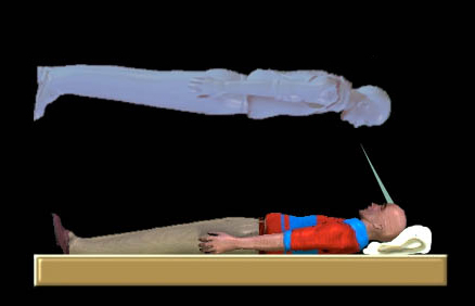
Out-of-body experiences; retrieved from Wikimedia Commons

1) Hallucinations: American psychiatrist and psychopharmacologist, Rick Strassman, has posited that the pineal gland releases DMT (N,N-dimethyltryptamine) into the dying brain [7]. This compound is known to exist in mammalian brains, as well as in many tropical plants, which are used by Amazonian shamans to elicit profound hallucinations and spiritual experiences. While there is no direct evidence that humans endogenously release DMT, the correlation between NDEs in CD survivors and NDEs in individuals who consume DMT in shamanistic rituals is striking and warrants further study. An alternative explanation that doesn't involve DMT is that disinhibition within the cerebral cortex may result in hyperactivity of N-methyl-D-aspartate (NMDA) receptors, which are known to produce hallucinations when stimulated by ketamine, a veterinary anesthetic [5].


2) Tunnel vision: This visual feature has been experimentally induced in pilots flying at high G-force, known as hypotensive syncope, which results from decreased blood flow to the eye. Neurons within the retina, specifically retinal ganglion cells (RGCs), are notoriously energy-hungry. These cells may therefore begin transducing aberrant electrical signals to the visual cortex (a part of brain responsible for decoding these signals into meaningful imagery) shortly after the onset of CD. Additionally, the retina is composed of distinct cell populations that process either central or peripheral information. It is plausible that this cytoarchitectural feature underlies the “tunnel vision” experienced in NDEs. CD-induced oxygen deprivation of RGCs have in fact been proposed as a mechanism for tunnel vision [8].


3) Serenity and Oneness: It is known that the dopamine and opioid systems are activated in the human brain under times of extreme duress and threat to survival. This represents an adaptive mechanism whereby pain and discomfort can be suppressed to facilitate the “fight-or-flight” response. Cardiac arrest likely qualifies as such a stressor, and it is known that blissful states result from endogenous opioid release. The cortex, or outermost part of the brain, is also farther from the main arterial conduits of blood than deeper, subcortical structures and may therefore be the first to become hypoxic. The major reward region in the brain, a deep structure known as the striatum, receives “go” signals from neurons in the cortex. Disinhibited cortical neurons that project to the striatum may be transmitting more "go" messages, and therefore elicit a burst of dopamine in the striatum, leading to a blissful state [6].


4) Life reviews: Rapid eye-movement (REM) sleep is a period of intense, vivid dreaming, memory consolidation, and physical paralysis. As the reader may attest, dreams within this relatively short (90-120 min) window seem to last for many hours. Mobbs and Watt, in their review of NDEs, cite the case of a diabetic patient in hypoglycemic shock who entered a sleep-like state with REM and later reported extensive life memories. They go on to suggest that because REM is enhanced by the neurotransmitter noradrenaline, putative release of this molecule in CD may drive vivid memory recall.


5) Disembodiment: It has been shown that stimulation of a part of the brain involved in proprioception (awareness of one’s physical body in space), known as the temporo-parietal junction (TPJ), can induce out-of-body experiences [9]. This region is also required for long-term memories that rely on proprioceptive information [10]. In line with these findings, CD patients may fail to properly integrate sensory information their bodies arriving at the TPJ. As a result, disruptions in self-representation may occur and precipitate a full-blown OBE.


Despite these creative and somewhat plausible arguments in favor of a neurobiological basis for near-death and out-of-body experiences, a fierce debate persists in the academic literature (for a full review, see [11]). This makes sense, as they're entirely conjecture and most of the data are entirely anecdotal. One highly contentious issue is over the reliability of the rare quantitative data presented in the field: electroencephalogram (EEG) recordings taken from CD patients. These repeatedly reveal a complete absence of brain activity, despite the vivid NDEs retold by CD survivors. If the brain were truly quiescent during vivid, complex NDEs, it would be necessary to completely abandon the neurobiological theory of consciousness as an emergent property of brain activity. Obviously, most researchers object. They tend to refute the EEG data, claiming that EEG is not sensitive enough to detect low-level activity that may persist for days in the anoxic (O2-deprived) brain [11]. To their credit, EEG only detects activity in the outermost brain tissue (the cortex), so deeper structures (like the striatum) may be full of undetectable electrical signaling. A more sensitive and powerful imaging modality is needed to provide clear answers.


Perhaps the most problematic issue lies with veridical reports from CD survivors with OBEs [12]. How can there by any biological explanation for a patient’s accurate visual recall of events occurring inside and outside of their exam room? This presents another serious challenge to the scientific materialist worldview. Most are quick to discount such experiences as flukes, conjured up in delirium, contorted by the grateful to fit their religious predilections. How will the scientific community treat the doctors, nurses, family members, and patients who make such claims (see Dr. Eben Alexander’s “Proof of Heaven”)? Do researchers and funding agencies have an ethical obligation to investigate them wholeheartedly?


One medical scientist, Dr. Sam Parnia, MD, PhD, thinks so and is attempting to empirically assess the veracity of stories from CD survivors with OBEs (listen to a recent NPR interview with Dr. Parnia). Dr. Parnia is principal investigator of the AWARE study (AWAreness during REsuscitation), which began in 2008. This five-year study involves 25 hospitals across Europe and North America, and its initial findings will be released later this year. Methodologically speaking, Dr. Parnia’s team places visual targets near the ceiling that could only be visible by someone reading it from above (hopefully for them, the disembodied CD patient). Whatever the AWARE study reveals, the fields of NDE, neuroscience, and medicine require scientists, clinicians, and the public to retain an objective viewpoint and an open mind. When new data emerge (yes, anecdotes are data too) that challenge scientific consensus, there is an ethical imperative to be skeptical, but not dismissive. Perhaps Sherlock Holmes offered the most prescient guidance on all of this when he said, “It is a capital mistake to theorize before one has data. Insensibly, one begins to twist facts to suit theories, instead of theories to suit facts.”


References

1. Moody RA. Life after life. Bantam Press; 1975.

2. Parnia S, et al. A qualitative and quantitative study of the incidence, features, and etiology of near death experiences in cardiac arrest survivors. 2001. Resuscitation. 48(2): 149-56.

3. van Lommel P, et al. Near-death experiences in survivors of cardiac arrest: a prospective study in the Netherlands. 2001. Lancet. 358: 2039-45.

4. Greyson B. Varieties of near-death experience. 1993. Psychiatry. 56: 390-99.

5. Mobbs D, et al. There is nothing paranormal about near-death experiences: how neuroscience can explain seeing bright lights, meeting the dead, or being convinced you are one of them. 2011. Trends in Cognitive Neuroscience. 15(10): 447-9.

6. Facco E, et al. Near-death experiences between science and prejudice. 2012. Frontiers in Human Neuroscience. 6: 1-7.

7. Strassman R. DMT, the spirit molecule: a doctor's revolutionary research into the biology of near-death and mystical experiences. Park Street Press; 2001.

8. Nelson KR, et al. Out-of-body experience and arousal. 2007. Neurology. 68: 794-5.

9. Blanke O, et al. The out-of-body experience: disturbed self-processing at the temporo-parietal junction. 2004. Neuroscientist. 11: 16-24.

10. McVea DA et al. Long-lasting working memories of obstacles established by foreleg stepping in walking cats require area 5 of posterior parietal cortex. 2009. Journal of Neuroscience. 29: 9396-404.

11. Braithwaite JJ. Towards a cognitive neuroscience of the dying brain. 2008. Skeptic. 21(2).

12. Holden JM, et al. Veridical perception in near-death experiences. Handbook of Near-Death Experiences. pp.185-211. Praeger; 2009.

Thursday, March 7, 2013

Live Neurons in Art: Components or Collaborators?

In his opening chapter of the biological art compendium “Signs of Life,” Eduardo Kac makes a particularly suggestive comment about the biological sciences in general.  I think this quote has even more significance to neuroscience specifically:


“The extreme difficulty in dealing with very complex biological interactions leads to the simplified treatment of life processes as quantified data that exhibit statistical patterns.  In turn, this can lead to an objectification of life and a disregard for the subjects and their rights.”[1]


From Zachary Weinersmith's

Saturday Morning Breakfast Cereal

This claim seems to echo Tom Wolfe's sentiment that scientific progress will lead to the death of the soul [2]: by reducing biological systems down to so many quantities and equations (all accurate within some statistical bounds), have we lost an important intuition about their intrinsic worth?  Is biology really just physical laws, with the same degree of moral importance as the law of gravity?  This reduction of the universe down to scientific law is called “naturalism,” and usually gets brought up during the discussion of free will or in religious contexts.  Here, though, I'd like to discuss naturalism in the context of what it means for the notion of “interests” in general- and consequentially, for ethical systems that are based on “interests.”


Fortunately, my favorite new-media artwork works as a stellar example of this issue.  In the Silent Barrage, a culture of rat neurons is used as part of an interactive robotic art installation.  Audience members walk among the robotic components (which are constantly drawing on sheets of paper), and are observed by overhead cameras.  The video feed from the cameras is translated into a pattern of electrical pulses which stimulate the culture, and the neurons within the culture respond by creating their own electrical activity.  This usually consists of bursts of activity that recruit the entire neural network (similar to epileptic seizures) which can be quieted through certain types of electrical stimulation.  Through the construction of this hybrid mixture of robotics and living neural tissue, the artists and scientists behind Silent Barrage give the audience the impression of walking through the mind of an artist suffering from epileptic seizures, in such a way that the interaction between the audience and the piece itself has the potential to silence these barrages of electrical activity.


The interaction between the artists and the biological tissue in Silent Barrage could potentially be evaluated as a sort of co-authorship: the human artists design the bulk of the piece, and the culture 'designs' (or perhaps 'performs' would be more appropriate) some of the details [3].  However, there are several differences between Silent Barrage and a traditional artistic collaboration.  First, the neural culture is without any sort of (forgive me) “cultural” understanding of the goals of the piece, lacking both the experience and capacity to understand the effect of it's actions in the hearts and minds of the audience.  Secondly, the neural component of Silent Barrage has no existence outside of the piece to pursue it's own interests - it is trapped inside the artwork, more similar to a raw material than a collaborator.


Left: Culture of rat neurons growing on top of a microelectrode array. Photo by Guy Ben-Ary.  Middle: Robotic drawing carriage used by Silent Barrage. Photo by Soyo Lee. Right: Drawing created by Silent Barrage- should the culture itself receive credit for this work?  Photo by Phil Gamblen.


Thirdly, and perhaps most importantly, any interests that the neurons of Silent Barrage have are being engineered to fill a particular need of the piece.  The electrical stimulation protocols used in the first showings of Silent Barrage were designed based on a simple relationship that had been previously observed [4]- the faster stimulation is applied, the less network wide bursting occurs. While living tissue (and the interests that such tissue might be said to have [5]) has been manipulated in several biological artworks [6] that preceded and followed Silent Barrage, unlike bacteria or osteoblasts, neurons are believed to make up the machinery that underlie the interests of morally (or at least legally) important beings- the rats who we aren't allowed to torture, the dogs we aren't allowed to neglect, and the humans that we aren't allowed to deny equality to.


The real issue here is that a system that looks suspiciously like your brain is being engineered to act as part of an artwork.  If we can engineer the interests of a neural system to match our own, does that mean that those interests can be considered our own tools?  And if a neural systems' interests are not just coincidentally equivalent to our own, but designed to be equivalent to our own, are they anything more than tools?  Does it still make sense to call them separate interests, or just extensions of our own? And lastly, if we can somehow convince ourselves that the 'interests' of a biological system are simply extensions of our own interests, what possible moral repercussions could come of our using biological systems in any way we please?


Want to cite this post?


 Zeller-Townson, RT. (2013). Live Neurons in Art: Components or Collaborators? The Neuroethics Blog. Retrieved on


-->, from http://www.theneuroethicsblog.com/2013/02/live-neurons-in-art-components-or.html


[1] Kac, Eduardo. "Art that Looks You in the Eye: Hybrids, Clones, Mutants, Synthetics, and Transgenics." Signs of Life. Bio Art and beyond (2007): 275-286.

[2]Wolfe, Tom. "Sorry, but your soul just died." Hooking up (1996): 89-109.

[3] Hughes, R. "The semi-living author: post-human creative agency." Architecture and Authorship, T. Anstey, K. Grillner, and R. Hughes, eds.(London, Black Dog Publishing) (2007).

[4] Wagenaar, Daniel A., et al. "Controlling bursting in cortical cultures with closed-loop multi-electrode stimulation." The Journal of neuroscience 25.3 (2005): 680-688.

[5] Varner, Gary E. In nature's interests?: interests, animal rights, and environmental ethics. Oxford University Press, USA, 2002.


[6] Catts, Oron and Zurr, Ionat. Tissue Culture and Art Project. Web 27 Feb 2013  <http://tcaproject.org/>

Friday, March 1, 2013

When the government can read your mind

We are now at a point where we can scan brain activity with fMRI, decode the patterns, and use the information to “read minds” or predict what a person is experiencing. For example, the Gallant Lab at the University of California, Berkley, published a paper in 20111 showing that by recording subjects watching a set of movies, they can estimate what visual features parts of the brain are encoding. Then, when they show you a new movie, their model can predict what you are seeing based on your brain activity.


This “mind-reading” has limitations: the reconstruction is primitive, worse on abstract or rare stimuli, and each subjects must be scanned many times to tune the model to his or her individual brain. However, this experiment proves the principle that we create models that use brain activity to predict dynamic conscious experience even with the low visual and temporal resolution and indirect measures of an fMRI. Other labs are progressing in different domains, for example Chang and colleagues were able to decode auditory cortex to reconstruct the individual words heard by subjects2.


This sort of technology has many implications, and provokes many questions—both technical and ethical:


  • Will the techniques extend beyond perception and allow us to read someone’s thoughts?

  • Could they be used to create a perfect lie director (or at least one orders of magnitude better than the polygraph)?

  • If such technology hypothetically existed, how would we use it?

  • How could we balance its potential benefits with potential for abuse?


fMRI for thought-reading Whether this approach of recording, modeling, and decoding will work with thoughts will depend on how similar our inner monologue is to actual audition. If our mental imagery uses the same pathways as sensory experience, and is encoded in a similarly, then we are not far away from it. However, this technique is unlikely to be able to extract thoughts the subject doesn’t want to share—it requires putting the subject in an fMRI (so could not be done surreptitiously), it requires compliance on the training taks, and even then inner-monologue can be consciously controlled.


fMRI as a Lie Detector Other groups are looking at lie detection—trying to use fMRI as a more accurate polygraph test. They work on the principal that certain patterns of brain activity will reflect the higher-order processing needed to suppress the truth. There are currently at least two private companies trying to market fMRI as a Lie Detector: Cephos Corporation and No Lie MRI. The sensitivity and specificity of these tests have not been well characterized.


Ethical use of thought-reading or lie detectors—Balancing benefits with risks In what ways is it appropriate to use these devices? Some are clearly beneficial, for example thought-reading may allow communication with otherwise inaccessible paralyzed “locked-in” patients.


Many possible uses are more ambiguous:


  • Could a theoretical thought-reading device ethically be used in questioning a criminal suspect, enemy combatant, or terrorist?

  • Could a person be apprehended and held responsible for thoughts they had if they had not yet committed a crime?

  • Is it ethical or legal for businesses to use lie-detectors in hiring, especially if there are of questionable efficacy?

  • At what level of accuracy (if ever) should a lie-detector would it be admissible in court?

  • Would results counted as testimony or biological evidence?


Even if we decide that some of these uses could be beneficial if used properly, how will we ensure they are used responsibly? Who will store the data produced and regulate that it will only be used for what it is meant to be?


These technologies will always be limited by the accuracy of our memories and our capacity for self-deception. Psychology studies have repeatedly shown malleability of memories and that subjective confidence isn’t a good correlate of accuracy. Even if a person thinks something is true and a lie detector verifies it, there is no insurance that it really is true.


As technology continues to improve, we must inform the greater public about what is currently possible, discuss these difficult questions, and make sure that our laws ensure the rights we want protected.


--Ben Kuebrich


Want to cite this post?


Kuebrich , B. (2012). When the government can read your mind. The Neuroethics Blog. Retrieved on

-->, from http://www.theneuroethicsblog.com/


Works Cited


1. Nishimoto NS, Vu AT, Naselaris T, Benjamini Y, Yu B, Gallant JL. (2011) Reconstructing Visual Experiences from Brain Activity Evoked by Natural Movies, Current Biology, 21(19): 10.1016/j.cub.2011.08.031


2. Pasley BN, David SV, Mesgarani N, Flinker A, Shamma SA, et al. (2012) Reconstructing Speech from Human Auditory Cortex. PLoS Biol 10(1): e1001251.doi:10.1371/journal.pbio.1001251